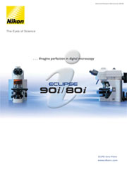
Nikon Eclipse 80i Advanced Research Microscope
Nikon Updated: 2007-06-19Microscopy and digital imaging combine to produce picture-perfect results.
Designed to truly harness the power of Nikon's CFI60 optics, the Eclipse 80i delivers remarkably high signal to noise ratios, producing fluorescence images that reveal more than ever before. Never has there been a research microscope so optimized for digital microscopy. The state-of-the-art design includes a vast array of advanced features, including Nikon’s proprietary “fly-eye” technology, VC Plan Apo objectives and both intelligent and motorized digital imaging heads for the ultimate digital imaging microscope system.
Unparalleled S/N ratio and contrast during fluorescence imaging
The digital imaging head and the universal epi-fluorescence illuminator incorporate Nikon’s unique Hi S/N Fluorescence System. The Noise Terminator directs stray light out of the detection path, delivering a signal-to-noise (S/N) ratio five times higher than our previous fluorescence system, increasing the image contrast during fluorescence microscopy and further extending the detection-level limit.
Excitation Balancer continuously adjusts excitation wavelength
During observations or imaging of multi-stained fluorescence specimens, the operator can easily emphasize a specific wavelength of the excitation light without changing the filter cubes. Allowing the observed intensity of each channel to be balanced against the intensity of the other channels when utilizing a multi-band filter set. This is accomplished by varying the distance the Excitation Balancer (optional) is slid into the excitation light path.
Six-filter Turret
The filter turret can accommodate six easily exchangeable filter cubes. Individual filter or mirror combinations in the cubes can be easily modified to create custom wavelength solutions. Phosphorescent filter labels are used on the turret cover, making it easy to see the names and positions of filter cubes in darkened rooms.
Upgraded DIC performance delivers uniformly crisp images with high contrast and resolution
The composition of the material used in the DIC prism has been changed to make it possible to obtain high-contrast DIC images with excellent resolution and uniform coloration at any magnification.
- Only two DIC condenser modules (dry) are necessary to allow observations at 10x-100x magnifications.
- Three different sheer types are available on our DIC prisms, allowing for standard, high-contrast or high-resolution observations.
- The shading (3D effect) of the image is controlled by simply rotating the polarizer on the base of the microscope (deSenarmont method) which eliminates the need to continually reach above the nosepiece as required on other systems.
Optics ideal for digital imaging
A revolutionary “fly-eye” lens array has been incorporated into the transmitted-light illumination optics to achieve highly uniform illumination, making loss of light intensity at the peripheries of the view field a thing of the past. Uniform brightness is possible at all magnifications, while completely filling the objective back aperture.
Plan Apo VC objectives deliver high-resolution images
Plan Apo VC (Violet Corrected) objectives have improved chromatic corrections to the periphery of the field of view to create superior digital images with the absolute highest resolution and uniform brightness – a feature perfect for stitching captured images. These objectives are highly recommended for confocal microscopy because of their high numerical aperture and axial chromatic correction even at the H-Line (405nm).
Digital-imaging head creates an optimum digital-imaging platform
This all-in-one digital-imaging unit for taking high-contrast, crisp fluorescence images integrates Hi S/N epi-fluorescence illumination with the Noise Terminator, dual-port beam-splitting module with zoom optics and binocular-eyepiece tube. A “Motorized Excitation” shutter control is provided.
Automatic detection of microscopy status
When the Nikon DS-Fi1 digital camera is mounted on the digital-imaging head, imaging data such as the objectives, imaging port, zoom magnification and fluorescence filter selection are automatically detected and can be saved as a text file in the image folder or output to an external imaging system. This eliminates the need to input data manually. Creating a large database of images taken at different settings is now easier than ever.
Optical Zoom function
A 0.8x-2.0x optical-zoom mechanism at the rear port allows imaging at desired magnifications. Unlike digital zoom, optical zoom produces smooth clear images because it allows users to match the optical resolution of the microscope system to the resolving power of the digital camera.
Dual Port
Two output imaging ports allow a variety of imaging devices to be mounted simultaneously. The front port is perfect for confocal and quantitative measurement applications as minimum lenses are used.
CFI60 Infinity Optics
The objectives of Nikon’s acclaimed CFI60 infinity optics have a 60mm parfocal distance, resulting in longer working distances and high NA’s, while producing crisp, clear images with high contrast and minimal flare. A flexible upgrade path is available to accommodate various intermediate modules.
Solid construction enables high-precision focusing
Utilizing computer-aided engineering (CAE), Nikon has significantly increased the stability of both the stage 'Z' movement and the arm section compared with previous Eclipse models. The increased stability minimizes the chance of unwanted blur or image shifts that can occur during high-magnification observations.
Stay-in-position stage handle
The handle of the new mechanical stage stays at a fixed position near the focusing knob throughout the full range of X/Y stage movement, so the operator’s hand can remain comfortably on the desk at the same position. The height and tension of the stage handle are adjustable.
Centering rotatable stage model
The rotatable-stage model allows image documentation at the desired angle, improving composition. Specimens that are sensitive to the optical axis orientation, such as DIC, can provide improved contrast and detail when rotated.
Ergonomic Tube
The new ergonomic binocular-eyepiece tube can be inclined at angles from 10° to 30° and the eyepieces can be extended up to 40mm. This ensures an optimum eye point and comfortable viewing posture, regardless of the operator’s physique or if intermediate modules have been attached.
A C-mount digital camera can be attached to the ergonomic tube using the optional DSC port with a 0.7x magnification.
Eye Level Riser
The eye-level riser can raise the eye point height in 25mm increments (up to 100mm maximum).
Product Brochure
Accessories Brochure
Related Manuals
Nikon Eclipse 90i Advanced Automated Research Microscope System
Nikon Eclipse FN1 Fixed Stage Microscope for Electrophysiological Research
Nikon Eclipse TE2000 Inverted Research Microscope Systems
Nikon Eclipse TE2000-PFS Inverted Research Microscope With Real-Time Focus Correction
Nikon NT88-V3 Micromanipulator Systems
Nikon Laser TIRF System
Nikon Eclipse 55i Biological Microscope
Nikon Eclipse TS100/TS100F Inverted Routine Microscope
Nikon BioStation IM Live Cell Recorder
Nikon Eclipse E200 Biological Microscope
Nikon Eclipse E100 Biological Microscope
Nikon COOLSCOPE Digital Microscope
