Nikon Eclipse 55i Biological Microscope
Nikon Updated: 2007-06-19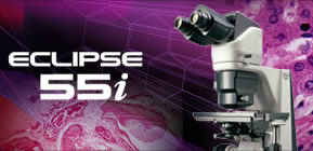
An advanced clinical microscope engineered for sophisticated observations over long hours of use.
The versatile Eclipse 55i clinical microscope offers powerful optical capabilites, unsurpassed ergonomic features and is optimized for digital imaging. Its unique white LED illumination system provides a homogenous and bright distribution of illumination across the field for optimal viewing and digital imaging, resulting in spectacular, vivid, true-to-life colors. Intuitive ergonomic features, like the stay-in-position stage handle and tilting/telescoping ergonomic eyepiece tube ensure ideal viewing posture, enabling long hours of observations to be carried out in perfect comfort.
White LED Illumination
Utilizing white LED illumination, the 55i is an ideal choice for brightfield applications. The color temperature remains constant even when the brightness is altered, making adjustiments of the color balance filter unnecessary. Due to its minimal power consumption, the 55i has an extremely long lamp lifecycle and the illuminator does not generate heat. An optional battery is available which allows the 55i microscope to be used anywhere in the world.
Renowned Nikon CFI60 Optics
Highly acclaimed optics combine the CF design with infinity optics utilizing a 60mm parfocal distance. The results are objectives with longer working distances and high NA's, producing crisp, clear images with minimal flare. The CFI60 optics are perfect for both observations through the eyepiece as well as capturing images with a digital camera. CFI60 optical design provides a flexible upgrade path to accommodate various accessories to be added to the microscope to meet individual applications.
Optional DSC Port
An optional DSC port can be combined with an ergonomic tilting/telescoping tube to balance user needs for both digital-image capture and comfortable viewing. Two models are available: a 0.7x lens that is designed to optimize the image to the 2/3-inch CCD, and a 1.0x lens that is designed to optimize the image to a 1-inch CCD. By matching the correct DSC port magnification to the CCD, the same area that is viewed through the eyepieces can be captured with a C-mount digital camera. A centering and focus adjustment mechanism is also provided.
Rock-solid Stability
Utilizing computer-aided engineering (CAE), the new microscopes boast superb durability and stability, even during applications in which they are upgraded with various attachments.
Ergonomic Eyepiece Tube
The ergonomic eyepiece tube can be inclined from 10 degrees to 30 degrees and the eyepieces can be extended 40mm. This ensures optimum eye point and a comfortable viewing posture, regardless of the operator's physique or if intermediate modules are used.
Refined Stage with Stay-in-position Stage Handle
Refined stage has a stay-in-position stage handle. The stage handle stays at the same position regardless of stage X-Y position. Because the stage handle and the focusing knob are always situated close to each other, users can easily and smoothly operate the controls with their hand resting comfortably on the desk. The height and torque of the stage handle are adjustable to further enhance comfort during operation. Alumite, a new hardening treatment, has been applied to the stage surface to increase durability and smoothness. This facilitates the smooth exchange of specimens while preventing the surface from being scratched by the repeated exchange of glass slides.
Easy Access Controls
Frequently used controls and switches for adjusting the field diaphragm and illumination intensity have been concentrated in the lower part of the right-hand side to minimize the operator's hand movements and enable operation without having to take your eyes off the specimen.
Ergo-View Cytodiagnosis Unit
The Ergo-View cytodiagnosis unit has been developed for easier and more comfortable cytology examinations, allowing fast and accurate motorized magnification changeover with hand switch. A unique quiet, vibration-free mechanism for magnification change ensures superb parfocality of images with no deviation in focusing, and easy slide marking while observing the specimen through eyepieces. Quick exchange of slides with one hand is possible by using an optional specimen holder for one slide.
Refocusing Stage
The stage can be lowered by pushing the lever down, and will return to its original position when the lever is pressed again. This eliminates the need to refocus the image manually each time the specimen is changed and the slide oiled, greatly improving microscopy productivity.
Pathology Tests
As the 55i uses white LED illumination, it can maintain the same color temperature even if the brightness is changed. Observations extending to many hours can be carried out in a natural posture without physical strain because an ergonomic tube enables the flexible adjustment of the eye point.
Cytodiagnosis Tests
The Erog-View cytodiagnosis unit makes magnification switching and slide marking faster and easier. With the 55i's unique white illumination, changes in the amount of LED light are aligned with magnification changes, enabling observation at a constant brightness.
Epi-fluorescence Microscopy
A dedicated turret-type epi-fluorescence illuminator has a quick-change mechanism combined with a unique filter-lock system and front-mounted shutter, providing the ultimate combination of performance and convenience in clinical fluorescence diagnostic microscopy.
Eclipse 50i/55i Accessories
Eclipse 50i/55i Brochure
Related Manuals
Nikon Laser TIRF System
Nikon Eclipse TS100/TS100F Inverted Routine Microscope
Nikon BioStation IM Live Cell Recorder
Nikon NT88-V3 Micromanipulator Systems
Nikon Eclipse TE2000-PFS Inverted Research Microscope With Real-Time Focus Correction
Nikon Eclipse E200 Biological Microscope
Nikon Eclipse TE2000 Inverted Research Microscope Systems
Nikon Eclipse E100 Biological Microscope
Nikon Eclipse FN1 Fixed Stage Microscope for Electrophysiological Research
Nikon COOLSCOPE Digital Microscope
Nikon Multizoom AZ100 Multi-Purpose Microscope
Nikon Eclipse 80i Advanced Research Microscope