Olympus FluoView 1000 Confocal Microscope
Olympus Updated: 2007-09-10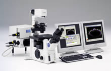
The Olympus FluoView™ FV1000 is a next-generation imaging system designed for high-resolution, confocal observation of both fixed and living cells. The FV1000 offers advances in confocal system performance while providing the speed and sensitivity required for live cell imaging with minimal risk of damage to living specimens.
In addition, the FV1000 offers a revolutionary synchronized laser scanning system called the SIM Scanner. While one laser stimulates, the second laser simultaneously provides high-resolution imaging. This coordination of laser stimulation and imaging makes the FV1000 an ideal choice for FRAP, FLIP and photoactivation.
SIM Scanner:
* Two independent laser scanners are incorporated in a single compact design for simultaneous laser stimulation and observation.
* The main scanner is used for high-resolution confocal imaging, while the second scanner is used for simultaneous laser stimulation.
* With simultaneous confocal observation during laser stimulation, rapid cell reactions that occur during or immediately following laser stimulation can be accurately captured.
* The circular "Tornado" scan provides highly efficient photobleaching and photoactivation in contrast to standard raster-scan patterns.
* The SIM Scanner is ideal for a variety of applications, including FRAP, FLIP, photoactivation, photoconversion, uncaging, laser ablation, and many others.
Spectral Scan System:
* Original system design features two independent spectral detection channels, each configured with a diffraction grating and variable slit for high-resolution wavelength separation and high-speed bandwidth selection.
* High-resolution spectral fluorescence detection in increments as low as 1nm enables accurate spectral unmixing of overlapping fluorescence emission signals. High-speed spectroscopy can be performed at 1msec/100nm.
* Two modes, Normal and Blind, are provided for spectral separation of two fluorochromes with similar fluorescence emissions.
* Variable bandwidth selection is available for each spectral PMT channel through simple adjustment of each variable slit. Fluorescence detection and spectral separation can be maximized for each channel through adjustment of the variable bandwidth to match the fluorochrome's peak emission.
High Sensitivity:
* A newly designed, high-sensitivity detection system provides efficient fluorescence detection, important for low laser conditions that minimize damage to living cells.
* Ion deposition filters are employed for increased sensitivity and full wavelength coverage.
* High-sensitivity PMTs, selected for high efficiency, can be used in either an Analog Accumulation (AAC) mode or in a Hybrid Photon Counting Mode (HPCM), ideal for low light specimens.
* With high signal-to-noise detection, the system excels in quantitation and photometric analysis of low-light recordings while minimizing cell damage.
High Precision and High Speed:
* Precise control of laser intensity via an advanced laser feedback system provides stable laser excitation throughout the time course of live cell studies, a necessary feature for accurate fluorescence quantitation.
* Spectral system provides 2nm wavelength resolution.
* High-speed imaging at 16 frames per second.
* High-speed spectroscopy at 1msec per 100nm.
* Fully automated internal stepper motor with Z-resolution of 0.01 micron.
Configurations:
* The FV1000 is fully motorized and configured upon the Olympus IX2 and BX2 series microscopes. The fully automated Olympus microscope platforms can be interactively controlled through the FluoView software, with internal stepper motor Z-resolution of 0.01 micron.
* The system is designed for easy expandability, with an open configuration that facilitates attachment of imaging devices to the various imaging ports, such as those provided for auxiliary CCD cameras.
* XY scanning is performed with a pair of galvanometric mirrors, yielding a wide scanning range to a field number of 18. Optical zoom (up to 50x) is available, with a maximum pixel resolution of up to 4096 x 4096.
* Up to four high-sensitivity photomultiplier tubes (PMTs) can be incorporated directly within the confocal fluorescence emission light path for high sensitivity detection of the fluorescence signal.
* A fifth PMT detector, dedicated for transmitted light imaging, may be used for the simultaneous detection of high-resolution Brightfield or DIC images with which the confocal fluorescence images may be overlaid.
* The FV1000 can be configured with up to 5 detection channels:
4 fluorescence detectors + 1 transmitted light detector or
2 fluorescence detectors + 2 spectral detectors + 1 transmitted light detector
Scanning Modes:
* XY: Single confocal optical image. 360° Image rotation is available.
* XZ: Single cross section image that cannot be obtained with a conventional microscope. The cross section image may also be rotated 360° or drawn as a free line (free line-Z).
* XT: Single line image recorded over time for time-lapse analysis with high temporal resolution. The line may be rotated 360° or drawn as a free line (free line-T).
* Point scanning: Series of intensity changes at a single point within the image with the highest temporal resolution.
* Xë: Spectral wavelength-scan recorded across a single line. The line may be rotated 360° or drawn as a free line (free line-ë). Spectral system required.
* XYZ: Series of confocal optical XY images recorded through the thickness of the sample.
* XYT: Single XY confocal optical image recorded over time, at an interval that can be arbitrarily chosen. Permits observation and analysis of live cell kinetics, such as changes in intracellular calcium, pH, etc.
* XYë: Spectral wavelength-scan for 2-dimensional XY confocal optical image. 360° Image rotation is available. Spectral system required.
* XëT: Spectral wavelength-scan across a single line recorded over time for time-lapse analysis. The line may be rotated 360° or drawn as a free line (free line-T). Spectral system required.
* XëZ: Spectral wavelength-scan recorded across a single XZ cross-section image. The cross section image may also be rotated 360° or drawn as a free line (free line-Z). Spectral system required.
* XYZT: XYZ series recorded over time, permitting observation and analysis of 3-dimensional changes over time in live cells.
* XëZT: Spectral wavelength-scan across a single XZ cross-section image recorded over time for time-lapse analysis. The line may be rotated 360° or drawn as a free line (free line-T). Spectral system required.
* XYëT: Spectral wavelength-scan for 2-dimensional XY confocal optical image recorded over time for time-lapse analysis. 360° Image rotation is available. Spectral system required.
* Automated Scanning Stage: Multi-location image acquisition is available for most imaging modes, including XYZ, XYT and XYZT. Requires the addition of an optional automated scanning stage.
Laser Choices:
* Multi-line Argon (457nm, 488nm, 514nm) laser
* 473nm Diode Laser
* Green Helium Neon (543nm) laser
* 559nm Diode laser
* 643nm Diode laser
* 440nm Diode laser
* 405nm Diode laser
TIRFM Illumination Unit:
* TIRFM illumination unit is available for the IX81 inverted microscope configured FV1000.
* The FV1000 with TIRFM unit and a CCD imaging system allow observation of high S/N imaging near the cell surface.
* The collimation and incident angle of the excitation laser toward the specimen is automatically set through FV1000 software .
* Laser lines can be switched via external TTL signals from the CCD imaging system.
Zero-Drift Compensator:
* Zero-Drift Compensator (ZDC) prevents focus drifts during long time-lapse observations.
* The FV1000 software activates ZDC before each scanning.
* A 785nm focusing laser minimizes fluorescence photo bleaching and photo-toxicity for specimens.
Scanning Stage Control:
* Scanning Stage Control software and a scanning stage allow Multi-Area Time-Lapse imaging of live cells.
* Observation parameters such as Zoom, PMT voltage, XYZ coordinates can be set for each area.
* Compatible with Well-plates as well as glass-slides and 35mm dishes.
FluoView 1000 Microscope Video (19.7 MB)
General Biological Brochure (PDF) (3MB)
FluoView 1000 Brochure (PDF) (1.3MB)
FluoView-LD405(E) (PDF) (1.39MB)
UIS Fluorescence Mirror Units Technical Specs (PDF) (448KB)
Related Manuals
Olympus FluoView 300 Confocal Microscope
Olympus Fluoview FV1000 MPE Multiphoton Microscope
Olympus IX81 Motorized Inverted Microscope
Olympus DSU Spinning Disk Confocal
Olympus IX71 Inverted Microscope
Olympus DP25 Digital Camera
Olympus IX51 Inverted Microscope
Olympus DP71 Digital Camera
Olympus BXFM Focusing Module Microscope System
Olympus DP20 Digital Camera
Olympus DP30 Monochrome Digital Camera
Olympus BX51WI/BX61WI Fixed Stage Research Microscopes