Olympus FluoView 300 Confocal Microscope
Olympus Updated: 2007-09-10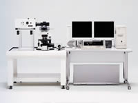
The Olympus FluoView 300 is a point-scanning, point-detection, confocal laser scanning microscope designed for biology research applications. Excellent resolution, efficiency of excitation, intuitive user interface and affordability are key characteristics of the Olympus FluoView 300.
The FluoView 300 configured permits simultaneous collection of up to 3 detection channels. The FV300 may be configured on either the IX2 inverted research microscope platform or the BX2 upright research microscope platform.
* Confocal microscopy can improve conventional fluorescence images by recording fluorescence generated from the focal plane within the sample, while rejecting all other light coming from above or below the focal plane. The efficient point-scan/pinhole-detection confocal optics of the FluoView systems virtually eliminate out of focus light to produce high-contrast images with superb resolution.
* The FluoView systems are fully integrated workstations that incorporate user-friendly image acquisition and image analysis software with high-resolution confocal optics that require no user alignment.
* An intuitive, Windows-based graphic user interface allows new users to quickly generate images in various scan modes, such as XY, XZ, XT, XYZ, XYT, and XYZT. Standard image formats, including TIFF and AVI, permit easy, direct export of FluoView images to off-line analysis packages.
* XY scanning is performed with a pair of galvanometric mirrors, yielding a wide scanning range to cover up to a field number of 20. The optical zoom (up to 10x magnification) can be performed by narrowing the scanning range while maintaining the maximum pixel resolution of up to 2048 x 2048 pixels.
* Automated Olympus microscope platforms can be interactively controlled through the FluoView software, with internal stepper motor Z-resolution of 0.01 micron.
* Fluorescence passes through a single confocal aperture, with five distinct aperture diameters available. Based on the objective magnification and numerical aperture, the optimal aperture diameter is indicated in the software.
* Brightfield and DIC images can be simultaneously recorded with the confocal fluorescence images. The transmitted light images, while non-confocal, are very useful when superimposed to the confocal image.
* Time lapse observation is possible on the FluoView with imaging rates of up to 4 frames/sec at 512 x 512, and line scan intervals at 0.5msec per line. Kinetic and ratiometric analysis is available for quantitation of fluorescence changes in live cells over time. Real time intensity plotting and ratiometric image display are possible.
* Multi-parameter experiments can be quickly designed and executed through the powerful PAPP (Programmable Acquisition Protocol Processor) software, an integrated component of the FluoView software. With the PAPP software, advanced time-lapse protocols involving photobleaching, photoactivation or multi-location acquisition can be easily generated and saved as routine protocols.
* TTL I/O signals can be generated to coordinate experiments timed with external instrumentation.
Scanning Modes Available:
* XY scanning: Acquires a single confocal optical image. 360° Image rotation is available.
* XYZ scanning: Acquires a series of confocal optical XY images through the thickness of the sample.
* XZ scanning: Acquires a single cross section image that cannot be obtained with a conventional microscope. The cross section image may also be rotated 360° or drawn as a free line (free line-Z).
* XYT scanning: Acquires a single XY confocal optical image over time, at an interval that can be arbitrarily chosen. Permits observation and analysis of live cell kinetics, such as changes in intracellular calcium, pH, etc.
* XT scanning: Acquires a single line image over time for time-lapse analysis with high temporal resolution. The cross section image may also be rotated 360° or drawn as a free line (free line-T).
* Point scanning: Acquires a series of intensity changes at a single point within the image with the highest temporal resolution.
* XYZT scanning: Acquires an XYZ series over time, permitting observation and analysis of 3-dimensional changes over time in live cells.
* Automated Scanning Stage: Multi-location image acquisition is available for most imaging modes, including XYZ, XYT and XYZT, with addition of an optional automated scanning stage.
Laser choices:
* Blue Argon (488nm) laser
* Multi-line Argon (457nm, 488nm, 514nm) laser
* Green Helium Neon (543nm) laser
* Red Helium Neon (633nm) laser
* Yellow Krypton (568nm) laser
* 442nm Helium Cadmium laser
* 440nm Diode laser
* 405nm Diode laser
Two high-sensitivity photomultiplier tubes (PMTs) are located directly within the FV300 confocal fluorescence emission light path for high sensitivity detection of the fluorescence signal. A separate, dedicated PMT may be used for the simultaneous detection of high-resolution Brightfield or DIC images with which the confocal fluorescence images may be overlaid.
FV300 optical path. Up to three channels of detection may be imaged simultaneously. Images may be scanned in any pixel array size up to 2048 x 2048, with each image digitized to 4096 gray levels (12-bit), permitting observation of fine image detail. Images may also be acquired in an automated sequential mode in order to reduce spectral cross talk between channels for multi-color images.
A single confocal aperture located directly within the emission light path is comprised of 5 distinct user-selectable sizes. The FluoView 300 software indicates appropriate aperture selection based on the magnification and numerical aperture of the objective in use.
A broad range of visible lasers, including the Argon (458nm, 488nm, 515nm), Green Helium Neon (543nm), Yellow Krypton (568nm) and Red Helium Neon (633nm), are available to suit most standard fluorescence applications. Each laser line may be independently regulated through software-controlled neutral density filter wheels or through an acousto-optical tunable filter (AOTF) for simultaneous or automated-sequential collection of multi-channel images. A choice of the 440nm diode laser and the 442nm Helium Cadmium laser is available for CFP/YFP FRET applications, providing optimal excitation of CFP and spectral separation from the YFP.
The 405nm diode laser is also available for most UV imaging applications, including nuclei imaging with DAPI. Unique laser modulation of the 405nm diode laser permits high-speed control of the light intensity, which can be important for photoactivation and photobleaching studies. The exceptional light stability of the 405nm diode laser is also important for time-lapse studies of living cells.
Flexibility in the selection of the microscope base, laser lines and optical filters permits researchers to configure the FV300 system for their specific research needs.
General Biological Brochure (PDF) (3MB)
FluoView 300 Brochure (PDF) (2.28MB)
FluoView-LD405(E) (PDF) (1.39MB)
UIS Fluorescence Mirror Units Technical Specs (PDF) (448KB)
UIS Objectives for Life Science (PDF) (359KB)
Related Manuals
Olympus DSU Spinning Disk Confocal
Olympus FluoView 1000 Confocal Microscope
Olympus Fluoview FV1000 MPE Multiphoton Microscope
Olympus DP25 Digital Camera
Olympus IX81 Motorized Inverted Microscope
Olympus DP71 Digital Camera
Olympus IX71 Inverted Microscope
Olympus DP20 Digital Camera
Olympus DP30 Monochrome Digital Camera
Olympus IX51 Inverted Microscope
Olympus BXFM Focusing Module Microscope System
Olympus DP12 Digital Camera