Hitachi HI VISION 8500 Ultrasound System
Hitachi Updated: 2008-09-03 RSS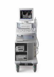
Powerful, flexible and fast, the HI VISION™ 8500 diagnostic ultrasound scanner combines leading edge technologies with user-oriented operation for exceptional imaging and functionality. The integrated SONO MR package consists of advanced techniques for image enhancement and control, including HI COMPOUND multi-angle spatial compounding, Wideband Pulse Inversion, tissue harmonics, HI RES adaptive imaging technology and Raw Data Freeze. These features can be selected separately or in various combinations to optimize B-mode, Color and Doppler images.
The Hi Vision 8500 also offers Sono IQ one-touch image optimization, real-time archiving with on-the-fly recording, a DVD rewriteable drive making even large dynamic clips manageable, and SonoElastography. Available exclusively on the HI VISION 8500, SonoElastography provides a completely new perspective on the physical properties of tumors and masses by determining and displaying the relative stiffness of tissue.
With leading edge features and options, including a wide variety of specialty and application-specific transducers, the HI VISION 8500 can be tailored to meet almost any clinical requirement.
HI VISION 8500 Features/Options
Ease of Operation
* Sono IQ one-touch image optimization
* Lightweight ergonomic probes
* Tri-color keyboard lighting for intuitive operation
* One touch access to commonly used functions
* User defined programmable keys
* Instantaneous key and control response
* Enhanced Cine and M-mode recording with Cine-max memory
* Powerful Built-in Measurement and Equation Editor
* Customizable Presets and Preset-Operation Functions
* Built-in hard disk, magneto-optical, floppy, and DVD drives
Advanced Performance
* HI COMPOUND Imaging
* HI RES adaptive Imaging
* Raw Data Freeze for Image Optimization in Freeze
* Tissue Doppler Imaging
* Octal Beam Processing for Fast Frame Rates
* 12 bit Gigasampling A/D for Precise Signal Reproduction
* Windows XP? based for reliability, stability, and familiarity
* HI VUE circuitry for streamlined operation and signal flow
* 4 Modes of dynamic Tissue Harmonic Imaging (dTHI)
o Includes Wideband Pulse Inversion Imaging (WPI)
* Directional Color Flow Angiography
* Real-time Doppler Measurements
* 3RD Generation Color Artifact Suppression
* Patient and Image Database Management System
o Includes storage space for 20,000 ~ 60,000 images
Optional Image Enhancements
* Real-time archiving
* SonoElastography imaging option
* WideView panoramic imaging option
* 3D imaging options (built-in or stand-alone)
* Steerable CW Doppler option
* ECG biophysical display option
* (dCHI) dynamic contrast harmonics imaging option
* Stress Echo examination option
* Pentax EUS and Fujinon Mini-probe options
* Dual Omni-directional M-Mode display option
Specifications
* 120Vac (100 ~ 240Vac selectable)
* 1.5kVA Power Consumption
* External Dimensions: (W) 22 x (D) 40 x (H) 59
* Weight: 350 lbs.
* DICOM 3.0 Store, Print, Worklist (Optional)
HI VISION 8500 Image Gallery
Related Manuals
Hitachi HI VISION 900 Ultrasound System
Hitachi HI VISION 6500 Ultrasound System
Hitachi HI VISION 5500 Ultrasound System
Olympus Nortec 1700 Conductivity Gauge
Olympus Sonic 260 Ultrasonic Multiplexer
Olympus Mini-Wheel Encoder
Olympus MCDU Motor Controler Unit
Olympus TRPP 5810 Remote Pulser/Preamplifier
Olympus Chain Industrial Scanner
Olympus HSTC-X01 Industrial Scanner
Olympus HST-X04 Industrial Scanner
Olympus HSMT-X03 Industrial Scanner