Olympus Fluoview FV1000MPE SIM Multiphoton Laser Scanning Microscope
Olympus Updated: 2009-03-27 RSS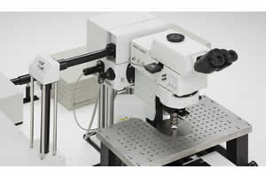
A Multiphoton Laser Scanning Microscope excellent for deep tissue imaging.
Olympus' Fluoview FV1000MPE is a multiphoton laser scanning microscope that allows fluorescence imaging deep within specimens. Utilizing pulsed IR lasers, the Olympus FluoView MPE is able to image hundreds of microns into a specimen thanks to the penetration of IR light and our long working distance objectives. The FluoView-MPE provides cutting-edge technology for various areas of scientific research such as neuroscience and cell biology.
Feature & Benefits
FV1000MPE SIM
* Visible imaging and IR stimulation
- No IR imaging
- No visible stimulation
* A less complex optical path with only two alignment points
* Newly designed delivery system for optimal performance with use of the Spectra Physics MaiTai DeepSee Laser
* The Olympus DeepSee lasers are specifically tuned to match Olympus' optical requirements
* 2 or 4 Channel Non-descan Detectors
* BX2 channel transmitted detector designed for high sensitivity detection for both transmitted fluorescence and SHG (on the upright platform only)
* Compact Design – one of the smallest footprints for MPE
- 1500mm X 1500mm (60in X 60in)
* Visible imaging available – 405nm, 440nm, 473nm, 458nm, 488nm, 515nm, 559nm, and 635nm laser lines
* Upright or inverted platforms
* AOM standard
* Dispersion compensation standard
Brochure
Related Manuals
Olympus Fluoview FV1000MPE TWIN Multiphoton Laser Scanning Microscope
Olympus Fluoview FV1000MPE Basic Multiphoton Laser Scanning Microscope
Olympus FluoView FV10i Self-contained Confocal Laser Scanning Microscope
Olympus LCV110 VivaView FL Incubator Fluorescence Microscope
Olympus DP72 Digital Camera
Leica DMI3000 B Inverted Microscope
Leica DMI4000 B Inverted Microscope
Leica DMI6000 B Inverted Microscope
Leica DMD108 Digital Microimaging Device
Leica DM2500 MH Materials Microscope
Leica DM4000 M Research Microscope
Leica DM6000 M Research Microscope