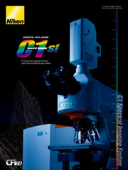
Nikon Eclipse C1si Confocal Microscope System
Nikon Updated: 2007-06-19A confocal system that combines the advantages of conventional fluorescence imaging and spectroscopy.
As research demands continue to grow, the need for greater detection of even more spectral information has become necessary, especially when discerning very similar or closely spaced spectral colors. The innovative C1si Confocal Microscope System gives you a level of flexibility, speed and spectral capability not available in conventional confocal systems, with detectors for both traditional fluorescence and spectral imaging in a wide range of applications.
Simultaneous acquisition of 32 channel spectral images
The C1si boasts a multi-anode PMT with 32 channels, the most of any confocal microscope. Innovations such as multiple high-speed digital conversion circuits and LVDS (Low Voltage Differential Signal) high-speed serial transmission technology allow a full 32 channels of spectral images to be obtained from a single scan. This allows for dramatically reduced imaging time and real-time observation.
One-scan acquisition of a broad 320 nm range
Three wavelength resolution settings of 2.5, 5, and 10 nm are available. At 10 nm, spectra over a full range of 320 nm can be obtained in a single scan, a capability unmatched by previous spectral imaging systems.
Gentle on Living Cells and Tissue
Spectral images over a broad wavelength range can be obtained with only a single laser excitation. Therefore, adjustment of laser intensity and PMT gain is simple and quick, and there is no need to make multiple scans to acquire a broad spectrum, keeping fluorescence fading and specimen damage at a minimum. The C1si spectral imaging system is gentle on living cells and tissue.
Acquiring Real Fluorescence Colors
Imaging of fluorescence spectra in real colors has been realized thanks to a host of new innovations for accurately correcting spectral data and wavelength resolution independent of pinhole diameter. Fluorescence spectra peak wavelengths and differences in spectral shapes can be detected by spectral acquisition with a high degree of reliability and accuracy. Whereas previously false colors were used to portray detail, the C1si allows observation of specimens in true color.
Peak wavelengths and spectral shapes obtained in C1si images closely match those obtained by the probe manufacturer.
Superb Error and Deviation Correction
Accuracy of spectra is maintained with highly precise correction technologies, including wavelength correction using emission lines and luminosity correction utilizing a NIST (National Institute of Standards and Technology) traceable light source. Also, multi-anode PMT sensitivity correction technology* allows correction of sensitivity error and wavelength transmittance properties on a per-channel basis, allowing researchers to minimize measurement errors and deviations among different equipment.
High Wavelength Resolution
High spectral resolution has been achieved through the use of a fine ruled diffraction grating supporting a 2.5nm channel width for high resolution confocal spectroscopy. Two other channel widths, 5nm and 10nm, are available for unmixing closely overlapping probes or for imaging four or more probes simultaneously.
Spectral detector with polarization Control Technology
Nikon’s proprietary DEES (Diffraction Efficiency Enhancement System) for polarization control has been adopted in the spectral detector of the C1si to maximize brightness. By co-aligning the direction of polarization, the efficiency of the diffraction grating is optimized, resulting in exceptionally bright images. In particular, increasing the diffraction efficiency in the long wavelength range leads to improved brightness and linearity of spectral data over the whole visible range from blue to red.
Multi-anode PMT
The spectral imaging detector utilizes a newly developed laser shielding mechanism. Coupled with high wavelength resolution that is independent of pinhole diameter, this mechanism prevents the reflected laser beam from contaminating data. The blocking mechanism can be moved freely with software, allowing users to block any laser wavelength, making the C1si compatible with virtually any laser selection.
High-efficiency Fluorescence Transmission Technology
The ends of the fluorescence fibers and detector surfaces use a proprietary anti-reflective coating to reduce signal loss to a minimum, achieving a high optical transmission.
Dual Integration Signal Processing
Newly developed DISP (Dual Integration Signal Processing) technology has been implemented in the image processing circuitry to improve electrical efficiency, preventing signal loss while the digitizer processes pixel data and resets. The signal is monitored for the entire pixel time resulting in an extremely high S/N ratio.
Unmixing of Fluorescent Signals without Crosstalk
The C1si software allows you to cleanly separate the signals of fluorescent probes, including those with closely overlapping spectra, such as CFP, RFP, YFP, and Alexa488. This is particularly useful in observations of multi-stained specimens with localized protein molecules, and in FRET experiments. Spectral separation of probe signals from autofluorescence is also possible.
Acquisition Features
Quick detector mode change — Switching from standard confocal imaging to spectral confocal imaging is a matter of turning the switch on the scanning head. The imaging mode of the EZ-C1 software is automatically switched.
Quick parameter setting — Each parameter of the spectral detector, e.g. excitation laser wavelength, wavelength resolution, or acquisition wavelength range, can be easily set from the menu with the mouse. When it is set, spectral imaging can be performed with common imaging procedures. Parameter profiles may be saved for later use. A binning function that combines signal from adjacent channels to increase brightness is provided. Therefore, when determining the target area, it is possible for users to lower excitation laser intensity and reduce damage to the specimen.
One-click acquisition of spectral images — Once spectral detector settings have been set, spectral confocal images can be acquired with a single click of the Start button.
One-touch fluorescence unmixing — Even without specifying a reference spectrum, simply drawing appropriate ROI’s (Regions of Interest) within the image and clicking the Simple Unmixing button allows separation of fluorescent probes based on their local spectra. Use the Unmixing button when you wish to specify the color each fluorescence probe will be displayed in after separation. The C1si contains a built-in database of spectral data provided from manufacturers of fluorescence labels, which can be specified as reference spectra for fluorescence unmixing. Users may also add spectral information derived from their own sample preparations into the database.
Unmixing red fluorochromes — Red fluorochromes, which had previously posed a challenge, are now simple to unmix as well.
Variable Color-Filtering
Wavelength information from the entire range can be obtained in a single spectral imaging operation. With C1si, there is no need to acquire a limited wavelength range and rescan the specimen to acquire the rest of the spectrum. After spectral imaging, you can easily display images that are filtered with any desired wavelength range.
Time-lapse recording of Spectral Images
Because all spectral channels are acquired in a single scan, C1s allows you to acquire temporally resolved spectral data. Time series acquisitions can be run at the maximum frame rate for the instrument, with a fixed delay, or on a user specified schedule.
Diascopic Observation
The C1si can acquire diascopic images simultaneously with either spectral or standard confocal images. Diascopic imaging techniques include DIC, brightfileld and phase contrast. This is especially helpful for locating fluorescent labels in tissues and cells.
FRAP Observation
FRAP (Fluorescence Recovery After Photobleaching) experiments are also possible on the macro program. The laser can be precisely pointed to photobleach a user defines ROI (Region of Interest) in a specific area part of the cell. The region can be a circle of elipse, a rectangle, or even a point or a line. Donut shaped regions can also be drawn allowing recovery of the bleached probe from pools located both inside and outside of the region. Other FRAP techniques including iFRAP (interval FRAP) and FLIP (Fluorescence loss in Photobleaching are also supported.
Simple, Flexible Microscope Controls
Switching between eyepiece observation and laser scanning modes is accomplished with a click on an icon. When the motorized TE2000-E Inverted Microscope or the ECLIPSE 90i Upright Microscope is used, the microscope can be controlled via the C1si system software, which frees users from the burden of changing optical paths and allows them to concentrate on data collection.
Eclipse C1si
Related Manuals
Nikon MM400/800 Industrial Measuring Microscopes
Nikon Eclipse C1 Plus Confocal Microscope System
Nikon NWL-641 IC Inspection Wafer Loader
Nikon LiveScan SFC Swept Field Confocal Microscope
Nikon SMZ1500 Zoom Stereomicroscope
Nikon NWL-860 IC Inspection Wafer Loader
Nikon Eclipse L200 Series IC Inspection Microscopes
Nikon SMZ1000 Zoom Stereomicroscope
Nikon Eclipse L300 Series FPD/LSI Inspection Microscopes
Nikon SMZ800 Zoom Stereomicroscope
Nikon SMZ645/660 Zoom Stereomicroscope
Nikon Eclipse LV150 Series Industrial Microscopes
