Nikon Eclipse C1 Plus Confocal Microscope System
Nikon Updated: 2007-06-19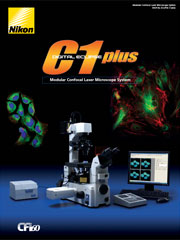
Multi-dimensional confocal fluorescence images with unsurpassed resolution and contrast.
The Eclipse C1 Plus is a modular confocal system that gives you the highest-quality digital imaging with an ultra-compact and lightweight design. The improved scan head optics of the C1 Plus boost optical performance to 400nm and its bi-directional scanner provides faster image acquisition. The system also includes support for X and Y scan axes rotation and new laser options for improved control over laser illumination intensity.
The Highest Quality Optical Performance
We've fused our first-class optics and electronics technologies to bring you a confocal system that provides state of the art resolution, contrast, and fluorescence image brightness.
A Wide Selection of Laser Types
The laser unit holds up to 3 different lasers, allowing you to use a variety of fluorescent probes. Also, changing filters to match the fluorescent dyes you're using is simple and fast. So as your research needs change, so will your microscope. A four laser unit is also available with direct modulation of diode lasers and acousto-optical tunable filter (AOTF) modulation of the other lasers. Laser choices include Argon ion, HeNe, Diode, and diode pumped solid state lasers (DPSS).
3-channel Simultaneous Detection
The C1 Plus supports almost any imaging technique you use today, including simultaneous 3-channel fluorescence, 3-channel plus DIC, time-lapse acquisition, and spatial analysis.
Ideal Instrumentation for FRAP
Fluorescence recovery after photo bleaching (FRAP) involves bleaching a targeted area of a cell with intense laser exposure and observing the recovery process. This is a useful tool for measuring the speed of molecular diffusion and movement. The C1 Plus can precisely target the laser exposure to the region of interest.
Advanced Scanning Features
The C1 Plus has a number of scanning features to make image acquisition simpler and faster. These include
Bidirectional Scanning: To improve image scan speed.
Scan Rotation: The image direction of long specimens, such as neurons, can be rotated without rotating the XY platform
Variable Delay in Time Lapse: Allows the intervals in time-lapse recordings to be varied.
Easy To Configure. Easy To Operate. Easy to Modify
Modular Components: Expansion and maintenance are easy.
Pre-Calibrated Modules: No need for calibration during set-up.
Compact Design: Does not fill up bench space. The C1 Plus features the world's smallest and lightest scanning head.
Intuitive Software: Most parameters can be modified with a single click of the mouse.
Four Position Pinhole Turret: Computer controlled selection of pinhole aperture allows customized balance between resolution and optical thickness.
TIRF Capabilities
The Nikon laser TIRF system can be used as a stand alone imaging device, in combination with widefield fluorescence, and/or in combination with a confocal microscope like C1-Plus or C1si. TIRF (total internal reflection fluorescence) microscopy improves signal to noise by exciting only those molecules located within a thin evanescent field, typically within 100nm or less of the coverslip. This technique complements confocal microscopy. The Nikon laser TIRF system mounts on the TE2000 in place of the standard fluorescence illuminator. It incorporates both TIRF and widefield fluorescence microscopy in a single device. Nikon has introduced two outstanding TIRF Plan Apochromat lenses with magnifications of 60x and 100x and NA = 1.49. These are the highest numerical aperture oil immersion objective lenses available anywhere!
FRAP Capabilities
FRAP (Fluorescence Recovery After Photobleaching) experiments are also possible on the macro program. The laser can be precisely pointed to photobleach a user defined ROI (Region of Interest) in a specific area part of the cell. The region can be a circle of elipse, a rectangle, or even a point or a line. Donut shaped regions can also be drawn allowing recovery of the bleached probe from pools located both inside and outside of the region. Other FRAP techniques including iFRAP (interval FRAP) and FLIP (Fluorescence loss in Photobleaching are also supported.
Eclipse C1 Plus
Related Manuals
Nikon Eclipse C1si Confocal Microscope System
Nikon LiveScan SFC Swept Field Confocal Microscope
Nikon MM400/800 Industrial Measuring Microscopes
Nikon SMZ1500 Zoom Stereomicroscope
Nikon NWL-641 IC Inspection Wafer Loader
Nikon SMZ1000 Zoom Stereomicroscope
Nikon NWL-860 IC Inspection Wafer Loader
Nikon SMZ800 Zoom Stereomicroscope
Nikon SMZ645/660 Zoom Stereomicroscope
Nikon Eclipse L200 Series IC Inspection Microscopes
Nikon Eclipse 50i POL Polarizing Microscope
Nikon Eclipse L300 Series FPD/LSI Inspection Microscopes