Leica VT1000 S Vibrating Blade Microtome
Leica Updated: 2009-02-07 RSS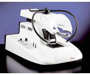
The Leica VT1000 S is a vibrating blade microtome, designed specifically to meet the highly sophisticated sectioning requirements in the fields of neurophysiology, neuropathology and experimental (brain) research – such as patch-clamping technique – and some industrial applications related to structural analysis of foam and other very soft materials.
The Leica VT1000 S can easily be adapted, depending on the application, by altering the frequency between 0 and100 Hz and by selecting the appropriate amplitude (0.2 to 1 mm in 5 steps). The instrument features a very fine adjustable knife advance speed between 0.025 to 2.5 mm/s and programmable sectioning window and specimen retraction.
Key Features
* Control of knife holder advance from 0.025 to 2.5 mm/sec
* Adjustable lateral knife holder amplitude of 0.2 - 1 mm and frequency (0-100 Hz)
* Automatic specimen advance in the single or continuous stroke mode
* Removable knife holder and buffer tray
* Ergonomic design
* Integrated cooling bath
* Section thickness totalizer
* Specimen retraction
* Orientable object plates
* Selection of accessories
The Leica VT1000 S vibrating blade microtome is designed to consistently produce thin sections of both fresh and lightly fixed tissues. Fresh nervous tissues, brain, spinal cord etc., are soft, fragile and extremely susceptible to mechanical damage.
Primary importance in the design of the VT1000 S was placed on accurate control of the knife or blade. To achieve this, a single edge razor blade or a sapphire knife is clamped into a holder that has an adjustable clearance angle of 5° - 15°. Accurate linear control of both advance speed and sectioning frequency is emphasized. Knife advance is now fully adjustable from 0.025 to 2.5 mm/sec Sectioning frequency has a range of 0 - 100 Hz, with an amplitude range of 0.2 to 1 mm in 5 steps.
Tissues are mounted directly onto a specimen plate, using cyanoacrylate adhesive, and mounted into a double walled buffer tray which is filled with physiological buffer. The buffer serves two purposes: it maintains the tissue in a living state and also provides a flotation medium for the sections. Both the knife holder and buffer tray are easily removed to ensure that there is no risk of reagent carryover or contamination when sectioning fresh or fixed tissues.
A programmable cutting window combined with accelerated return knife speed ensures rapid sectioning of even the smallest specimens.The superior sections produced with the VT1000 S contain a significant number of viable cells and are invaluable for electrophysiological studies on the living cell.
User Manual
Certificates
Related Manuals
Leica LN22 Nitrogen Freezing Device
Leica VT1200 Vibrating Blade Microtome
Leica VT1200 S Vibrating Blade Microtome
Leica Q550 IW General Imaging Workstation
Leica Q550 MW Materials Workstation
Leica AF6000 E Entry Level Fluorescence Microscope
Leica AF6000 Advanced Fluorescence Imaging System
Leica AF6500 Fluorescence Station
Leica AF7000 Dedicated Fluorescence Live Cell Station
Leica MM AF Integrated System
Leica AM TIRF MC Microscope
Leica DCM 3D Measuring Microscope